Research

Optical methods for the brain and for cancer

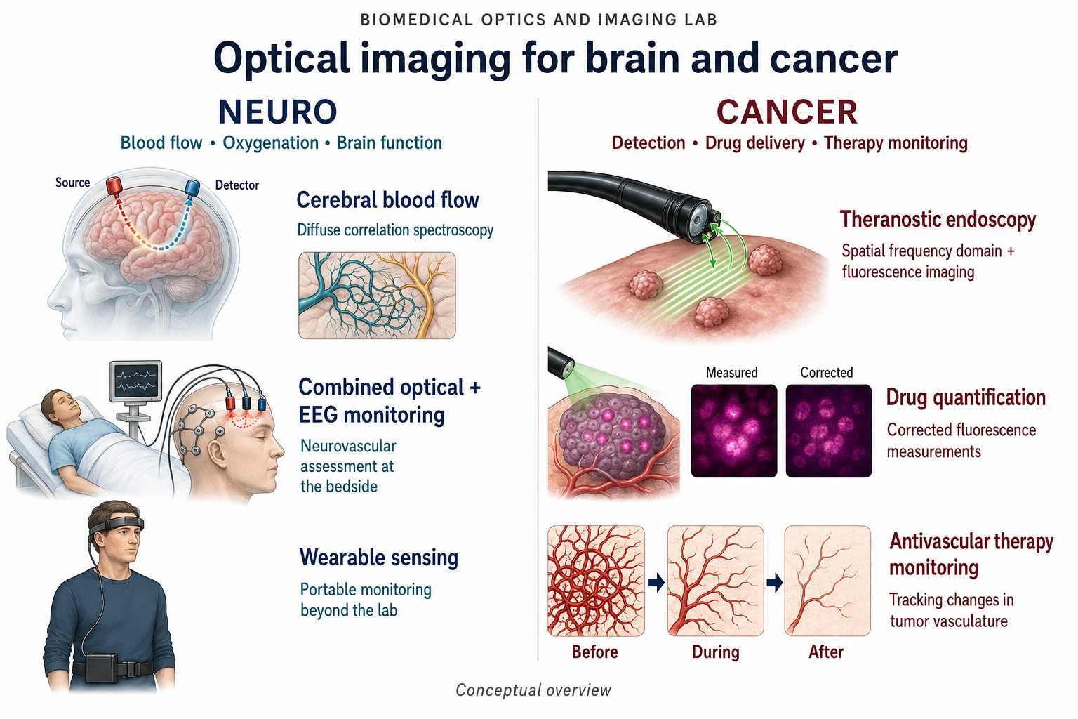
The lab develops noninvasive in vivo imaging and spectroscopy techniques for diagnosis and treatment in these 2 areas, and takes them through to instruments that work outside the optical bench.

Overview of the 2 branches of the lab's work. Neuro covers blood flow, oxygenation and brain function, shown as cerebral blood flow measured by diffuse correlation spectroscopy with a source and a detector on the head, combined optical and EEG monitoring at the bedside, and wearable sensing outside the lab. Cancer covers detection, drug delivery and therapy monitoring, shown as theranostic endoscopy using spatial frequency domain and fluorescence imaging, drug quantification comparing a measured against a corrected fluorescence image, and antivascular therapy monitoring showing tumor vasculature before, during and after treatment.

Neuro research

Measuring cerebral blood flow, oxygenation and their slow oscillations at the bedside, without a contrast agent and without moving the patient.

Time gated diffuse correlation spectroscopy for brain function

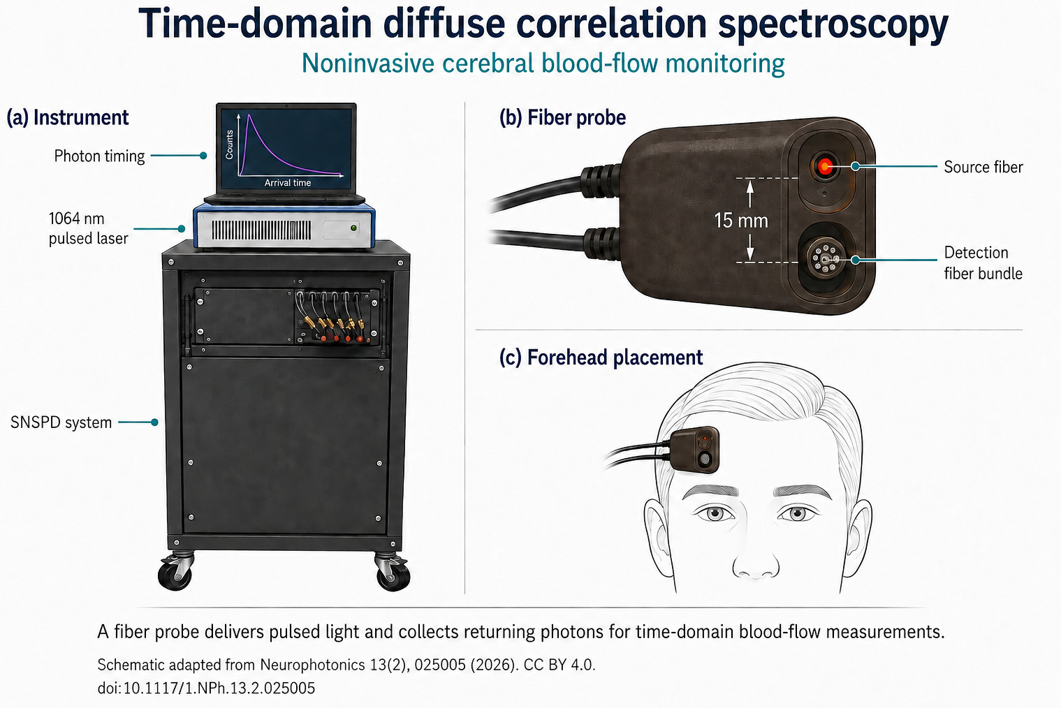
Diffuse correlation spectroscopy measures blood flow by reading the way speckle intensity fluctuates as near infrared light passes through moving red blood cells. The difficulty in the brain is that most detected photons never reached the cortex, so the signal is dominated by scalp and skull.

This project addresses that by gating on photon arrival time. Late arriving photons have travelled further and carry more cortical weighting, so separating them from early arrivals recovers a depth resolved measurement. Working at 1064 nm with superconducting nanowire single photon detectors improves tissue penetration, photon throughput and the safety margin under exposure limits at the same time. The applications are neurointensive care and functional neuroimaging.
Time gated photon detection, in 2 panels. Panel a shows a source and a detector on the surface with a labelled source to detector separation. Early photons follow a shallow path through scalp and skull, late photons follow a deeper path into the brain, so early arrivals carry greater superficial sensitivity and late arrivals greater brain sensitivity. Panel b plots detected photon counts against photon arrival time, with an early gate marked near the peak of the distribution and a late gate marked on its tail.

Characterizing traumatic brain injury

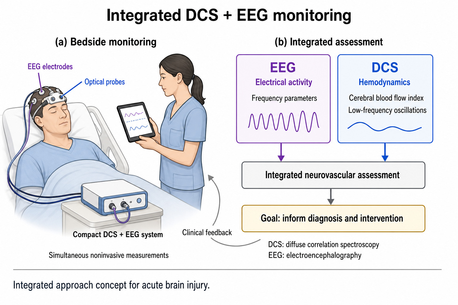
Electroencephalography and optical spectroscopy see different halves of the same event. EEG reports electrical field potentials, optical measurement reports cerebral blood flow and oxygenation. Recorded together and noninvasively, they give a fuller view of the neurovascular unit than either does alone.

The project uses that combination to characterize brain injury and to test whether low frequency oscillations in perfusion carry information that neither modality shows on its own. Work so far indicates that oscillation measures shift after injury more clearly than blood flow alone does, which makes them a candidate early biomarker of neurovascular disruption.

Integrated diffuse correlation spectroscopy and EEG monitoring, in 2 panels. Panel a shows a patient in a hospital bed wearing a cap carrying EEG electrodes and optical probes, cabled to a compact combined system, with a clinician reading traces on a tablet. Panel b shows EEG contributing electrical activity as frequency parameters and diffuse correlation spectroscopy contributing hemodynamics as cerebral blood flow index and low frequency oscillations. Both feed an integrated neurovascular assessment whose stated goal is to inform diagnosis and intervention, with clinical feedback returning to the bedside.

Cancer research

Quantifying how much drug has reached a tumor, and watching it work. Here an agent is part of the method, so the measurement has to be corrected back to an absolute concentration rather than reported as brightness.

Ovarian cancer detection and therapy

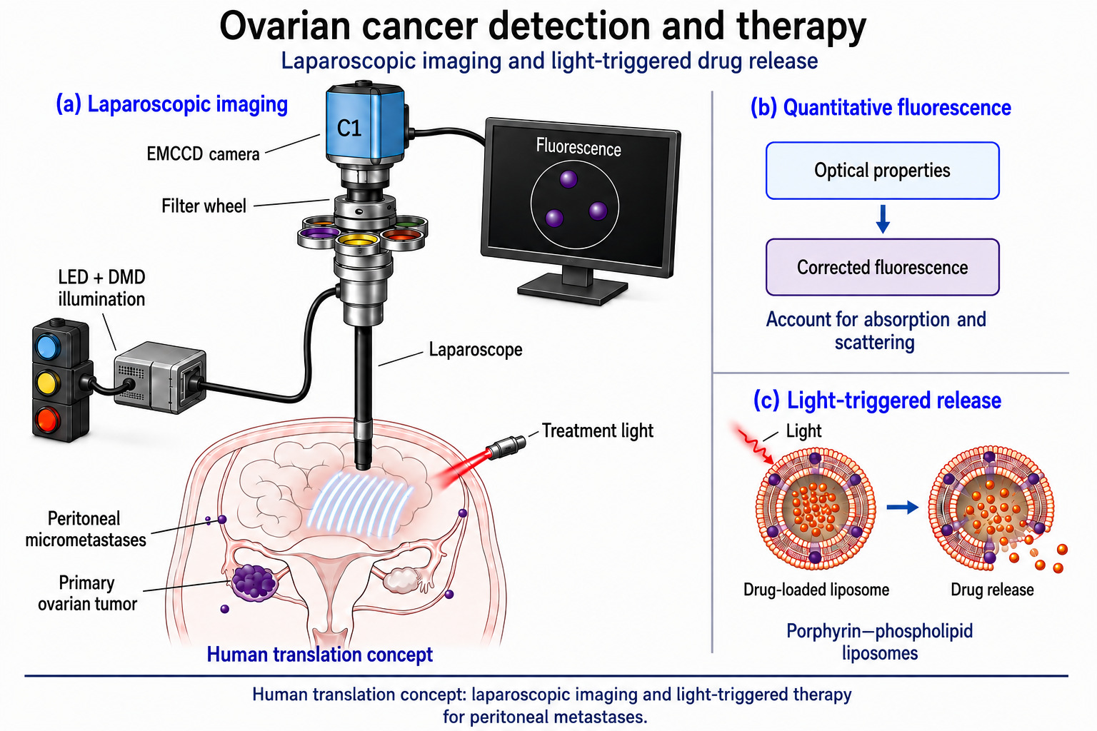
Peritoneal micrometastases are hard to see and often resist systemic therapy. This project develops quantitative fluorescence imaging through a laparoscope to detect them, and pairs the imaging with chemophototherapy, where near infrared light triggers release of a chemotherapeutic from porphyrin phospholipid liposomes at the target site.

Because tissue absorption and scattering distort any raw fluorescence measurement, the imaging is built on spatial frequency domain imaging, which recovers optical properties first and then corrects the fluorescence to an absolute concentration. That correction is what turns a picture into a dose, and it is what makes intraoperative monitoring of treatment plausible during minimally invasive surgery.

Ovarian cancer detection and therapy, drawn as a human translation concept, in 3 panels. Panel a shows a laparoscope in the abdomen projecting a striped pattern onto the peritoneal surface, with a primary ovarian tumor and scattered peritoneal micrometastases labelled, LED and digital micromirror illumination feeding the laparoscope, an EMCCD camera behind a filter wheel, a fluorescence view on a monitor, and a separate treatment light. Panel b shows optical properties feeding a corrected fluorescence measurement, to account for absorption and scattering. Panel c shows a drug loaded porphyrin phospholipid liposome releasing its contents when light reaches it.

Alongside

Fluorescence imaging and tomography

Time domain and continuous wave fluorescence molecular tomography, used to quantify tumor characteristics and to follow photochemical reactions such as photosensitizer photobleaching during treatment.

Photoacoustic imaging

Light in, sound out. Absorbed optical energy generates an acoustic wave, which gives access to blood vessels and oxygenation at high spatial resolution and without a contrast agent, at depths where purely optical imaging has lost its resolution.

Where the results are

Every claim on this page is set out in full in the peer reviewed literature. The publications page lists selected papers with links, and the complete record is on Google Scholar.